|
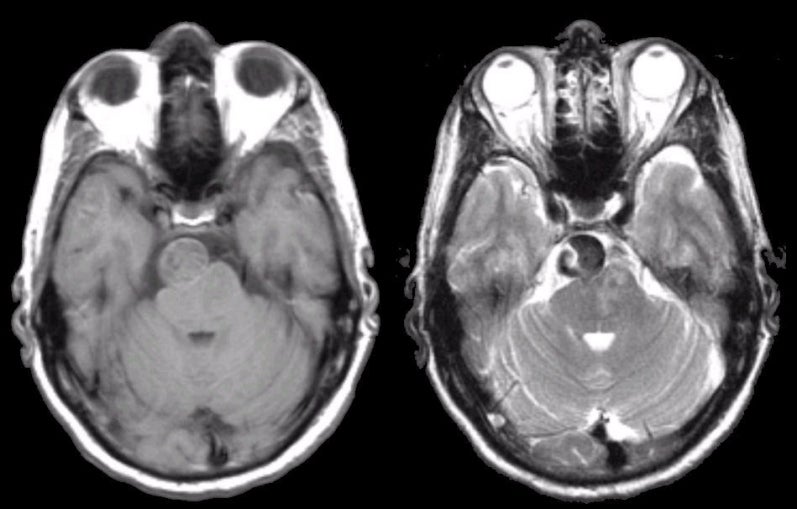
A 45 year-old woman developed headaches followed by a right hemiparesis and a right fourth nerve palsy. |
![]()
![]()
| Basilar Artery Aneurysm. Axial MRI scans of the brain. (Left) T1-weighted image; (Right) T2-weighted image. Note the circular mass compressing the upper pons. The circular mass is a fusiform basilar aneurysm. Although aneurysms typically come to clinical attention when they bleed, rarely they enlarge resulting in mass effect and focal neurological signs (in this case, the fourth nerve palsy). If one looks closely at the T2-weighted image, one can see abnormal signal in the left pons. This is an infarction of the basis pontis, the cause of the right hemiparesis. Presumably, a clot within the aneurysm occluded one of the small perforators to the pons resulting in the infarction. |
Revised
11/18/06.
Copyrighted 2006. David C Preston